top of page
Petrographic and Sample Preparation Services for Microscopy & Microanalysis
Maciej G. Sliwinski (Research Geologist, Ph.D)
Rock Thin Sections & Grain Mounts
Consultation Services: Chemostratigraphy of shales; surface-to-subsurface correlation
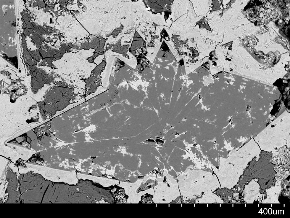
Gallery: Uroliths in thin-section
Cross-section of bladder stone composed of calcium carbonate and apatite, extracted from 12 year old dog. Comparative imaging by optical microscopy (PPL, XPL) and back-scattered electron imaging.
Veterinary care & stone extraction: Dr. Zenon Knasiak, DVM and Dr. Tomasz A. Wietecha, MD
Electron microscopy and x-ray analysis by Bill Schneider and John F. Fournelle (Electron Microbeam Research Labs, The University of Wisconsin-Madison).


Set 1 XPL (1000 µm scale)

Set 1 PPL (1000 µm scale)

Set 2 XPL (500 µm scale)

Set 2 PPL (500 µm scale)

Set 3 XPL (250 µm scale)

Set 3 PPL (250 µm scale)

Set 4 XPL (500 µm scale)

Set 4 PPL (500 µm scale)

BSE-SEM Image 1

BSE-SEM Image 2

BSE-SEM Image 3

BSE-SEM Image 4
BSE-SEM Image 5

BSE-SEM Image 6

Set 5 XPL (500 µm scale)

Set 5 PPL (500 µm scale)

Set 5 XPL (250 µm scale)

Set 5 XPL (250 µm scale)

Set 6 XPL (500 µm scale)

Set 6 XPL (250 µm scale)

Set 7 XPL (500 µm scale)

Set 7 PPL (500 µm scale)

Set 7 XPL (250 µm scale)

Set 7 PPL (250 µm scale)
 IMG_3146 (2) |  IMG-0259_edited_edited |
|---|---|
 Misia urolith 1 (Medium) |  T3_PPL_40x (Medium) |
 100X BSE Urolith 6 |
bottom of page